


MicroCT Scanner
Our Nikon XTH 225 X-Ray MicroCT Scanner and 3D Reconstruction system is similar to a medical CT scanner, but with higher voltage and resolution. It is used to investigate 3D structures on a fine scale, with a maximum resolution of 3µm. An x-ray source beams x-rays through a rotating object, collecting thousands of images, and then using a computer algorithm to combine them into a 3D model of individual "voxels" (3D pixels) of differing x-ray density.
Example projects include:
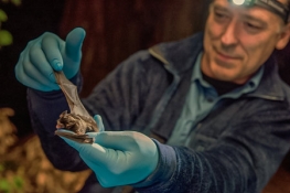
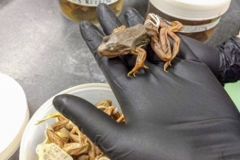
- Mapping the detailed three-dimensional anatomy of amphibian noses, to better understand their adaptation to land vs. water
- Developing new techniques for assessing sudden oak death oomycete infection of plants and associated soils
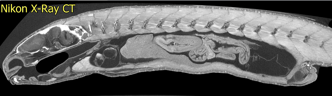
- Non-destructive reconstruction of fossil fish internal anatomy
- Understanding form and ecological function of the calcified structures of marine invertebrates by biomimetic modeling
- Resolving otolith structure in three dimensions non-destructively, which will allow for more sophisticated assessment of growth patterns across ontogenetic transitions
- Developing new ways to detect differences between cryptic species of bryozoans
- Studying three-dimensional diversification of bacular morphology among chipmunks, in order to assess the covariation of morphology and genetic relatedness
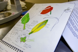
- Documenting the diversity of conifer seed-cone anatomy to better understand their diversification
- Comparing inner ear structures across the groups of cartilaginous fishes, to understand ecomorphological relations that can be extrapolated to interpret the ecology of fossil taxa.
The instrument was acquired with a grant from the National Science Foundation (NSF) Major Research Instrumentation (MRI) Program.